With appropriate training, physiotherapists can perform and interpret point-of-care ultrasound scans for the assessment of acute haemarthrosis to a level comparable to an experienced sonographer

© Shutterstock

graphic/j_jhp-2022-0008_fig_011.jpg

Haemophilia is a haematological condition with orthopaedic manifestations. People with haemophilia (PwH) are prone to several complications. Haemarthrosis is the most frequent complication, accounting for 70–80% of all bleeding episodes [1]. Although any joint may be affected, hinge joints, particularly the ankles, knees and elbows, are the most commonly involved [1]. Blood within the joint space has detrimental effects on all joint structures and leads to the development of haemophilic arthropathy [2,3]. A single haemarthrosis is capable of causing the same long-term arthropathy as seen in recurrent haemorrhages [4,5]. Time between the initiation of joint bleeding symptoms and treatment with factor replacement therapy is crucial; however, some haemarthroses may present ambiguously. On initial presentation it may be difficult to assess if acute joint pain is due to a joint bleed or underlying arthropathy [6]. Recent evidence suggests that clinical examination alone is not sensitive enough to detect small amounts of blood within a joint [4,6,7]. Therefore, each bleeding event requires early and complete bleed assessment and management to ensure the best possible outcomes for PwH.

Magnetic resonance imaging (MRI) is the gold standard for the detection of haemarthrosis, however it is expensive, often inaccessible, and may require sedation of children to ensure that the images are not compromised by patient movement [8]. Computed tomography (CT) is another sensitive method; however, the benefits of CT may not outweigh the downside of ionising radiation [8,9]. Ultrasound is time-efficient, nonionising, and relatively inexpensive [8,10,11,12]. Ultrasound can detect complex fluid suggestive of blood within the joints of patients who are clinically asymptomatic, leading to the recommendation that ultrasound be used in combination with the clinical exam to inform treatment decisions following haemarthrosis [7]. However, clinical integration of ultrasound is limited by timely access to sonographers/radiologists with knowledge and experience in haemophilia. Further, treatment of haemarthrosis is time-sensitive, and same-day diagnostic imaging appointments are not always feasible. Point-of-care ultrasound (POCUS) is a modality that has the potential to address many of these challenges. POCUS is performed by a health care professional (HCP) at the bedside or in the ambulatory clinic, in combination with the clinical examination to identify the presence or absence of a specific clinical finding [13]. POCUS should be utilised when time saving for diagnosis or treatment is critical to patient care [13]. However, POCUS is a highly user-dependent modality, and there is a risk of misdiagnosis if it is used to aid clinical decision-making by inexperienced or untrained HCPs [14].

Proficiency with the clinical examination and an understanding of the role of POCUS are important competencies for physiotherapists [15]. POCUS has been described within physiotherapy practice in orthopaedics or sport injuries to detect atrophy, tendon, ligament or muscle injury, in rheumatology to assist clinical decisions, and as a potential tool for physiotherapists working in critical care [16,17,18,19,20,21]. It is important that physiotherapists who are using POCUS have confidence in their interpretation and findings, as this could impact the credibility of the profession, patient safety, support from regulatory colleges and licensing bodies, and collaboration with medical colleagues. A survey of orthopaedic surgeons and radiologists in the Netherlands reported no additional value of physiotherapist-performed POCUS in primary care [22]. This is a single study that assessed the subjective opinions of survey respondents; perceived disadvantages of physiotherapist-performed POCUS were false-positive or false-negative results, lack of experience, inadequate training, and the inability to correlate the reported findings on POCUS with other forms of imaging [22]. Even though this study reported a low survey response rate and a potential for response bias, the lack of trust radiologists and orthopaedic surgeons reported for physiotherapist knowledge and performance of POCUS in primary care should be addressed through future studies [22]. Rathi and colleagues investigated the inter-rater reliability of glenohumeral joint translation using POCUS [23]. Although high intra-rater reliability (physiotherapist: ICC 0.86–0.98, expert sonographer: 0.85–0.96) was found, it was moderate to good for posterior measurements (ICC 0.50–0.75) and poor to moderate for anterior measurements (ICC 0.31–0.53). These results suggest that to improve inter-rater reliability with an expert sonographer, the physiotherapist may benefit from additional or a different form of training [23]. Similar findings were reported by Thoomes-de Graaf and colleagues, who found a kappa coefficient of 0.36 between physiotherapists and radiologists on the use of diagnostic ultrasound in patients with shoulder pain across four diagnostic categories [24]. Although the level of agreement was low, this study reported that physiotherapists with more experience and training had a higher level of agreement with the radiologist than novice physiotherapists [24].

Training appears to be an important contributor to inter-rater reliability of physiotherapist-performed POCUS. Mayer and colleagues found excellent inter-rater reliability (ICC range 0.76–0.97) between a physiotherapist, physiotherapy students, and an expert physician sonographer following eight hours of structured formal training as a group and a one-hour private practical training session with the expert sonographer [25]. An inter-examiner agreement study of physiotherapists in the Netherlands found an acceptable level of overall agreement (61.7–93.6%) and specific positive agreement (43.9–91.4%) for detecting rotator cuff tears and other pathology [26]. The physiotherapists in this study had obtained certification on basic musculoskeletal ultrasound skills and completed a six-hour training programme specific to the study protocol with an expert in musculoskeletal sonography [26].

Physiotherapists in haemophilia treatment centres (HTCs) have extensive knowledge of anatomy, pathophysiology, and functional implications of a bleeding disorder on the musculoskeletal system. A global survey of HTCs found that the majority (70%) of POCUS scans were completed by physiotherapists [27]. In this study, an interdisciplinary panel of haematologists/oncologists, radiologists, and physiotherapists reported that physiotherapists are appropriate users for the acquisition and interpretation of POCUS scans in HTCs [27]. While several researchers have studied diagnostic ultrasound and the correlation with disease activity and haemophilic arthropathy, inter-professional agreement and an evaluation of image quality for physiotherapist-performed POCUS in PwH with acute haemarthrosis has not been investigated [28,29,30,31,32,33]. Inter-professional agreement and evaluation of image quality are important measures of competency and acceptable use of POCUS. Image quality provides evidence to support the diagnosis of a bleed and decreases the chance of artifact incorrectly demonstrating pathology. Given the role of physiotherapists within HTCs in Canada, and the emergence of POCUS, the present pilot study aims to add novel research to this discussion.

Objectives:

  1. To determine the level of agreement between physiotherapist and sonographer-performed POCUS to assess for the presence or absence of blood in acute haemarthrosis in people with haemophilia A and B.

  2. To compare the quality of the ultrasound images obtained by the physiotherapist to those obtained by the sonographer.

METHODS

Study design

This study was a single blind, prospective, pilot study.

Participants

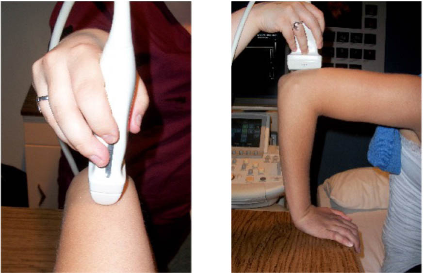
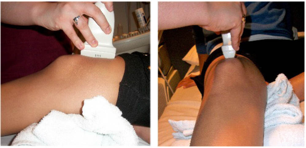
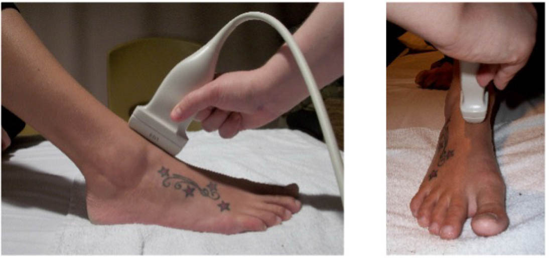
A convenience sample of PwH with a suspected acute hemarthrosis of the elbow, knee, or ankle were recruited from a single HTC in Canada. The physiotherapist (KS) who performed the POCUS scan is a member of the Canadian Physiotherapists in Hemophilia Care, successfully completed the McMaster University Mohawk College POCUS Training Program for Acute Hemarthrosis and Synovitis, and has 17 years of experience in haemophilia care. The training programme includes 12 hours of online didactic modules and a two-day, 12-hour practical training module with instructor-led hands-on practice [34]. The didactic modules include summative assessments, and the practical component includes an assessment of competency using a simulated performance environment. The assessments were created to model the Sonography National Competency Profile developed by Sonography Canada and the Sonography Canada Clinical Skills Assessment Tool for this specific application of POCUS [34]. The sonographer (LF) who performed the ultrasound scan is a senior sonographer in the diagnostic imaging department at a large tertiary care hospital and has over 30 years of clinical experience in sonography. A single radiologist (NS) with 12 years of experience in ultrasound imaging and 10 years’ experience in paediatric imaging, provided oversight to the study and reviewed all POCUS scans and case report forms.

Study procedures

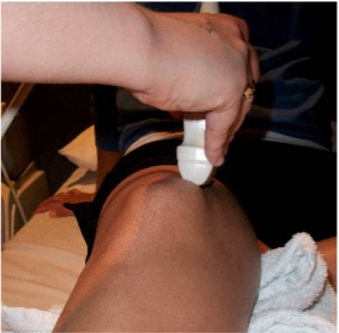
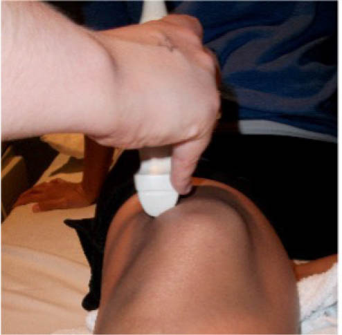
The study procedure consisted of a POCUS performed by a physiotherapist and a sonographer. The POCUS scanning procedure is presented in Appendix 1. The POCUS scans were performed in the haemophilia ambulatory clinic at patient presentation, one-week follow up, and two-week follow-up. The order of assessment was based on clinician availability. The sonographer was allowed to add additional images to the imaging protocol given their area of expertise, but the physiotherapist was instructed to acquire the images according to the scanning procedure. Ambiguous results were referred to the diagnostic imaging department for further formal investigation. Both the physiotherapist and the sonographer were blinded to each other's findings and to the results of previous scans. Methods of blinding included the use of a private clinic room and each clinician performing their assessment and documentation independently. Case report forms were placed in a sealed envelope. POCUS images were saved on the hard drive of the POCUS machine (GE Logiq) using an anonymous participant identification number.

Inclusion and exclusion criteria

Patients (age >1 year of age) with haemophilia A or B, with an acute haemarthrosis involving the elbow, knee, or ankle who presented to the clinic within five days of symptom onset were eligible to participate. Participants were excluded if there was an open wound over the scanning area, if an ultrasound scan of the haemarthrosis had already been completed, or if they were not able to read and understand English.

Outcome assessment

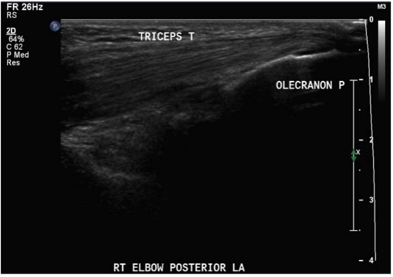
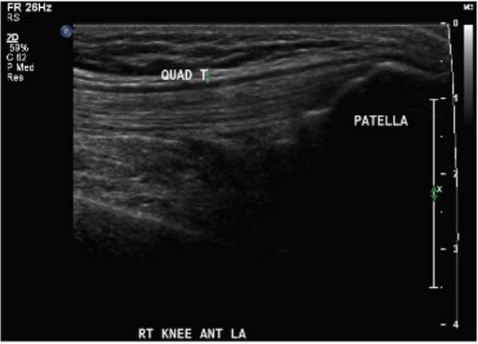
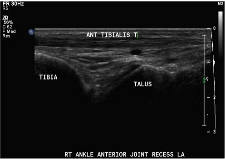
Outcomes were assessed at presentation, one-week follow-up, and two-week follow up, resulting in a three-week study period. For the primary objective, the outcome of interest was the binary decision on the presence or absence of blood within the joint. As the technique and protocol in this study was specific to haemophilia, the criteria used to distinguish blood from effusion on ultrasound was blood presents as a complex fluid collection with mixed echogenicity and displaceable speckles on real time compression and effusion presents as simple anechoic fluid with the absence of echoes [35]. In the context of haemophilia with no symptoms suggestive of infection, complex effusions with mixed echogenicity can be assumed to represent haemarthrosis based on previous studies that have documented the accuracy of this approach using joint aspiration [35]. The physiotherapist completed the scanning protocol and interpreted the findings to make the binary assessment. Since interpreting ultrasound falls outside the scope of the sonographer, the sonographer provided an impression on the presence or absence of blood on the case report form. The radiologist read the sonographer images and provided a final diagnosis. The radiologist also reviewed the images of the physiotherapist performed ultrasound. To compare the level of agreement, the radiologist's final diagnosis was compared to the physiotherapist's interpretation. Both the physiotherapist and the sonographer recorded inconclusive ultrasound findings as absence of blood within the joint.

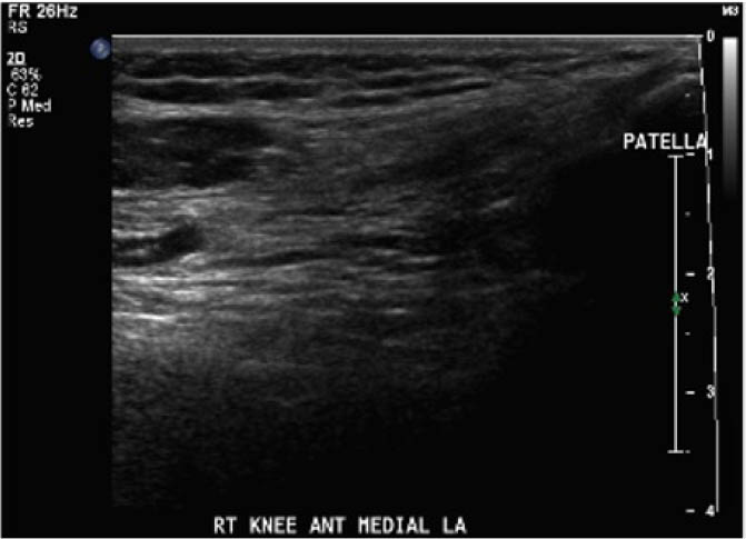
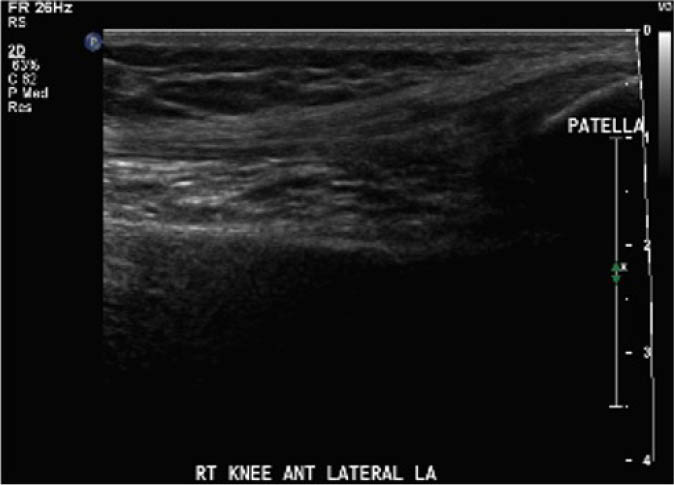
For the secondary objective, criteria used to evaluate image quality were appropriate pre-sets, depth, field of view, focus, gains/time gain compensation, colour and/or power Doppler, with suitable landmarks and annotation. Image quality was evaluated by the radiologist post hoc and rated as optimal, acceptable, or sub-optimal. Optimal was defined as good image quality with optimal ultrasound settings and correct annotation/documentation. Acceptable was defined as good image quality, with one image setting that should have been better optimized or a minor error in annotation/documentation that did not impact the interpretation of the POCUS scan. Sub-optimal was defined as poor image quality with more than one image setting not sufficiently optimised or an error in annotation/documentation that impacted the radiologists’ interpretation of the POCUS scan.

Statistical analysis

For the primary objective, the prevalence of positive findings was calculated. The inter-rater agreement of the binary assessment of the presence and absence of blood within the joint was assessed with the kappa coefficient and 95% confidence intervals for the total sample and interpreted according to the categories by Landis and Koch [36]. As this is a pilot study, we did not set an a priori threshold for agreement. Observed agreement, specific positive agreement and specific negative agreement were also calculated to provide the results in a clinically relevant format [37]. For the secondary objective, the quality of the images was independently rated by the radiologist. Descriptive statistics including counts and percentages of optimal, acceptable, and sub-optimal for the physiotherapist and the sonographer performed POCUS scans were reported.

Ethics approval

The proposed study received research ethics board approval from the Hamilton Integrated Research Ethics Board. All participants provided informed consent to participate in the study.

RESULTS

Thirteen PwH met the inclusion criteria and were recruited into the study. Two of the POCUS scans involved elbows (15.4%), five (38.5%) ankles, and six (46.2%) knees. The median age of participants was nine years (interquartile range: five years).

Level of agreement on the presence or absence of blood within the joint

As presented in Table 1, the kappa coefficient was k=0.80 (95% CI, 0.59–1.00). The prevalence of positive findings was 70.8%, observed agreement was 91.7%, the specific positive agreement was 94.1%, and the specific negative agreement was 85.7%. The sonographer was absent and unable to complete three POCUS scans, these scans were excluded from the level of agreement analysis.

Table 1

Level of agreement on the presence or absence of blood within the joint

KAPPA COEFFICIENT AGREEMENT

Overall (n= 36) k=0.80 (95% CI, 0.59–1.00) p=0.000 Prevalence: 70.8%
OA: 91.7%
SPA: 94.1%
SNA: 85.7%

[i] k: kappa coefficient; OA: observed agreement; SPA: specific positive agreement; SNA: specific negative agreement

Quality of ultrasound images

Post hoc analysis of the quality of the ultrasound images is shown in Table 2. The physiotherapist-performed POCUS scans demonstrated that 84.6% of the images were rated by the radiologist as optimal, 15.4% were rated as acceptable, and none were rated as sub-optimal. For the sonographer-performed POCUS scans, 88.9% of the images were rated as optimal, 11.1% were rated as acceptable, and none of the scans were rated as sub-optimal.

Table 2

Quality of ultrasound images

QUALITY OF ULTRASOUND IMAGES
OPTIMAL ACCEPTABLE SUB-OPTIMAL
Physiotherapist 84.6% 15.4% 0%
Sonographer 88.9% 11.1% 0%

[i] Optimal: good image quality with optimal ultrasound settings and correct annotation/documentation

[ii] Acceptable: good image quality, one image setting should have been better optimised or a minor error in annotation/documentation that did not impact POCUS interpretation

[iii] Sub-optimal: poor image quality with more than one image setting not sufficiently optimised or an error in annotation/documentation that impacted POCUS interpretation

DISCUSSION

Although pilot in design, this study adds to the emerging literature supporting the quality of physiotherapist-performed POCUS. The level of agreement between physiotherapist and sonographer is encouraging, suggesting that a trained physiotherapist is able to acquire and interpret POCUS scans of acute haemarthrosis in patients with haemophilia A and B at the same level of expertise as an experienced sonographer [36]. The specific positive agreement was greater than the specific negative agreement indicating better agreement when confirming the presence rather than the absence of blood within the joint. Clinically, these results indicate that if the physiotherapist performed and interpreted the POCUS scan as positive for presence of blood within the joint, the probability that the sonographer and radiologist would also confirm haemarthrosis is 94.1%. Encouraging results were also found for the absence of blood within the joint with the probability of absence of blood at 85.7%. While agreement was less for the absence of blood, the results were still high. Clinically, this supports physiotherapist consultation with radiology to determine whether further imaging is required if the POCUS scan indicates a lack of blood in the joint but other indicators such as patient symptomatology, mechanism of injury, inhibitor status, trough level, and underlying joint health, are all highly suggestive of haemarthrosis. The quality of the images obtained by the physiotherapist were optimal and comparable to the sonographer. This indicates that the trained physiotherapist was able to produce images that support the diagnosis on the presence or absence of a joint bleed with a low likelihood of imaging artifact incorrectly demonstrating or missing pathology.

In clinical practice, POCUS images are seldom stored for future review or comparison [38]. This process has been questioned as it limits the possibility of performing quality assurance audits and does not acknowledge the importance of reviewing serial scans to assess for the evolution/resolution of pathology [38]. To be consistent with this process and to maintain the independence of POCUS scans, the current study blinded the physiotherapist and the sonographer to the images and findings from previous scans. However, given the evolution of blood on ultrasound, the potential for underlying joint arthropathy in PwH, and the role of normal physiologic fluid in synovial joints, it may be important for the POCUS operator to have access to previous POCUS scans. Being able to access previous images can facilitate analysis of the clinical evolution of pathology and physiologic fluid, which may have implications on the level of agreement between the trained physiotherapist and sonographer as there may be variations in agreement at different stages of recovery. Also, recording previous images may decrease health care costs. If the POCUS scans are done with similar standards as diagnostic imaging, accessing stored images may avoid the need for repeat scans if clinical questions arise that may require consultation with radiology. These considerations may have implications for education and clinical practice and will be important areas for future study.

The current study had a number of strengths. The physiotherapist was trained to a set standard for this specific application of POCUS by an accredited academic institution. Both the physiotherapist and the sonographer were blinded and were provided with a standardised scanning protocol, with the order of assessment randomised based on clinician availability. Both the physiotherapist and the sonographer used the same ultrasound machine and after each POCUS scan the machine was returned to the main screen to maintain blinding and the independence of scans. All 13 participants recruited into the study attended all study visits. Lastly, the study procedures were consistent with the traditional pathway in diagnostic imaging. The study was designed in this manner to ensure that the same quality and standard of care was provided in the clinic setting.

Although this is a pilot study, its main limitations are the small sample size and inclusion of a single physiotherapist and sonographer, both of which may impact generalisability. While the results suggests that a short training programme provided the physiotherapist with an appropriate level of education and training in the performance, acquisition, and interpretation of POCUS scans in PwH, this needs to be replicated with physiotherapists and sonographers with varying levels of training and experience. It would be interesting to compare the competencies of sonographers with no musculoskeletal experience to physiotherapists who have completed POCUS training specific to the musculoskeletal system. Future inter-professional agreement studies should also consider including other members of the haemophilia comprehensive care team, such as physicians/haematologists and nurses, who may be using POCUS in clinical practice. In addition, with the decreasing annualised bleeding rates in PwH [39], a multi-centre trial would be needed to obtain a sufficient number of suspected bleeding episodes for a definitive study.

This study focused on hinge joints of the knee, ankle, and elbow, which account for the majority of haemarthrosis in PwH and are easily accessible with relatively simple POCUS scanning protocols. Future research would need to look at the inter-professional agreement and image quality for more complex joints, such as the ball-and-socket joints (i.e. shoulder and hip). Although haemarthrosis could occur in patients with other musculoskeletal injuries, these results should only be applied to the assessment of haemarthrosis in patients with haemophilia [40,41,42]. The protocol and training received by the physiotherapist in this study was specific to haemophilia and it is important to remember that one of the downsides of POCUS occurs when users extrapolate beyond their protocol and training [13]. Generalising the findings of this study to patients with other conditions should therefore be done with caution. However, this study does demonstrate that within a relatively short period of formal training, including both didactic and practical curricula, physiotherapists can become proficient in POCUS. Given their background knowledge in anatomy and physiology, this study lends support for physiotherapists to be trained to use POCUS with different patient populations and conditions.

CONCLUSION

Optimal image quality and an excellent level of agreement between the physiotherapist and sonographer-performed POCUS for the assessment of acute haemarthrosis in people with haemophilia A and B was observed. This pilot study found that a physiotherapist who received appropriate training in the McMaster University Mohawk College Training Program can perform and interpret POCUS scans for the assessment of acute haemarthrosis to a level that is comparable to an experienced sonographer. Further investigation is warranted.

ACKNOWLEDGEMENTS

Dr Alfonso Iorio and Dr Davide Matino for their support of the study procedures.

Wendy Lawson and the faculty at Mohawk College for their development and collaboration in the McMaster University Mohawk College Training Program for the Management of Hemophilia.

The authors would like to thank the participants in this study for their involvement.

Informed consent has been obtained from the participants in the study reported in this paper.

Notes

[5] Funding

Funding for this research was provided by an investigator-initiated grant from Pfizer Canada, the Health Professional Investigator Fund and the Clinical Health Professional Research Award at Clinical Health Professional Research Award at Hamilton Health Sciences, and the McMaster Children's Hospital Foundation.

[6] Conflicts of interest Conflict of interest

Anthony Chan is supported by Hamilton Health Sciences and the McMaster Children's Hospital/Hamilton Health Sciences Foundation Chair in Pediatric Thrombosis and Hemostasis.

Monica R. Maly is funded by The Arthritis Society and the Canadian Institutes of Health Research – Institute of Musculoskeletal Health and Arthritis. The funders played no role in the design, conduct or reporting of this trial.

REFERENCES

1 

Srivastava A, Santagostino E, Dougall A, et al. WFH Guidelines for the Management of Hemophilia, 3rd edition. Haemophilia 2020; 26 (Suppl 6): 1–158. doi: 10.1111/hae.14046.

A SrivastavaE SantagostinoA DougallWFH Guidelines for the Management of Hemophilia, 3rd editionHaemophilia202026Suppl 6115810.1111/hae.14046

2 

Dunn AL. Pathophysiology, diagnosis and prevention of arthropathy in patients with haemophilia. Haemophilia 2011; 17(4): 571–8. doi: 10.1111/j.1365-2516.2010.02472.x.

AL DunnPathophysiology, diagnosis and prevention of arthropathy in patients with haemophiliaHaemophilia2011174571810.1111/j.1365-2516.2010.02472.x

3 

Jansen NWD, Roosendaal G, Lafeber FPJG. Understanding haemophilic arthropathy: an exploration of current open issues. Br J Haematol 2008; 143: 632–40. doi: 10.1111/j.1365-2141.2008.07386.x.

NWD JansenG RoosendaalFPJG LafeberUnderstanding haemophilic arthropathy: an exploration of current open issuesBr J Haematol20081436324010.1111/j.1365-2141.2008.07386.x

4 

Melchiorre D, Linari S, Innocenti M, et al. Ultrasound detects joint damage and bleeding in haemophilic arthropathy: A proposal of a score. Haemophilia 2011; 17: 112–17. doi: 10.1111/j.1365-2516.2010.02380.x.

D MelchiorreS LinariM InnocentiUltrasound detects joint damage and bleeding in haemophilic arthropathy: A proposal of a scoreHaemophilia2011171121710.1111/j.1365-2516.2010.02380.x

5 

Rodriguez-Merchan EC. Cartilage damage in the haemophilic joints: pathophysiology, diagnosis and management. Blood Coagul Fibrinolysis 2012; 23: 179–83. doi: 10.1097/MBC.0b013e32835084dd.

EC Rodriguez-MerchanCartilage damage in the haemophilic joints: pathophysiology, diagnosis and managementBlood Coagul Fibrinolysis2012231798310.1097/MBC.0b013e32835084dd

6 

Ceponis A, Wong-Sefidan I, Glass CS, von Drygalski A. Rapid musculoskeletal ultrasound for painful episodes in adult haemophilia patients. Haemophilia 2013; 19(5): 790–98. doi: 10.1111/hae.12175.

A CeponisI Wong-SefidanCS GlassA von DrygalskiRapid musculoskeletal ultrasound for painful episodes in adult haemophilia patientsHaemophilia20131957909810.1111/hae.12175

7 

Aznar JA, Abad-Franch L, Perez-Alenda S, et al. Ultrasonography in the monitoring of management of haemarthrosis. Haemophilia 2011; 17(5): 826–8. doi: 10.1111/j.1365-2516.2011.02538.x.

JA AznarL Abad-FranchS Perez-AlendaUltrasonography in the monitoring of management of haemarthrosisHaemophilia2011175826810.1111/j.1365-2516.2011.02538.x

8 

Beyer R, Ingerslev J, Sorensen B. Muscle bleeds in professional athletes--diagnosis, classification, treatment and potential impact in patients with haemophilia. Haemophilia 2010; 16(6): 858–65. doi: 10.1111/j.1365-2516.2010.02278.x.

R BeyerJ IngerslevB SorensenMuscle bleeds in professional athletes--diagnosis, classification, treatment and potential impact in patients with haemophiliaHaemophilia20101668586510.1111/j.1365-2516.2010.02278.x

9 

Dauty M, Sigaud M, Trossaert M, et al. Iliopsoas hematoma in patients with hemophilia: a single-center study. Joint Bone Spine 2007; 74(2): 179–83. doi: 10.1016/j.jbspin.2006.05.014.

M DautyM SigaudM TrossaertIliopsoas hematoma in patients with hemophilia: a single-center studyJoint Bone Spine20077421798310.1016/j.jbspin.2006.05.014

10 

De la Corte-Rodriguez H, Rodriguez-Merchan EC. Treatment of muscle haematomas in haemophiliacs with special emphasis on percutaneous drainage. Blood Coagul Fibrinolysis 2014; 25(8): 787–94. doi: 10.1097/MBC.0000000000000159.

H De la Corte-RodriguezEC Rodriguez-MerchanTreatment of muscle haematomas in haemophiliacs with special emphasis on percutaneous drainageBlood Coagul Fibrinolysis20142587879410.1097/MBC.0000000000000159

11 

Balkan C, Kavakli K, Karapinar D. Iliopsoas haemorrhage in patients with haemophilia: results from one centre. Haemophilia 2005; 11(5): 463–7. doi: 10.1111/j.1365-2516.2005.01123.x.

C BalkanK KavakliD KarapinarIliopsoas haemorrhage in patients with haemophilia: results from one centreHaemophilia2005115463710.1111/j.1365-2516.2005.01123.x

12 

Querol F, Rodriguez-Merchan EC. The role of ultrasonography in the diagnosis of the musculo-skeletal problems of haemophilia. Haemophilia 2012; 18(3): e215–26. doi: 10.1111/j.1365-2516.2011.02680.x.

F QuerolEC Rodriguez-MerchanThe role of ultrasonography in the diagnosis of the musculo-skeletal problems of haemophiliaHaemophilia2012183e2152610.1111/j.1365-2516.2011.02680.x

13 

Chawla TP, Cresswell M, Dhillon S, et al. Canadian Association of Radiologists Position Statement on Point-of-Care Ultrasound. Can Assoc Radiol J 2019; 70(3): 219–25. doi: 10.1016/j.carj.2019.06.001.

TP ChawlaM CresswellS DhillonCanadian Association of Radiologists Position Statement on Point-of-Care UltrasoundCan Assoc Radiol J20197032192510.1016/j.carj.2019.06.001

14 

Solomon SD, Saldana F. Point-of-care ultrasound in medical education-stop listening and look. N Engl J Med 2014; 370(12): 1083–5. doi: 10.1056/NEJMp1311944.

SD SolomonF SaldanaPoint-of-care ultrasound in medical education-stop listening and lookN Engl J Med2014370121083510.1056/NEJMp1311944

15 

Rhon DI, Deyle GD, Gill NW. Clinical reasoning and advanced practice privileges enable physical therapist point-of-care decisions in the military health care system: 3 clinical cases. Phys Ther 2013; 93(9): 1234–43. doi: 10.2522/ptj.20120148.

DI RhonGD DeyleNW GillClinical reasoning and advanced practice privileges enable physical therapist point-of-care decisions in the military health care system: 3 clinical casesPhys Ther201393912344310.2522/ptj.20120148

16 

Whittaker JL, Teyhen DS, Elliott JM, et al. Rehabilitative ultrasound imaging: understanding the technology and its applications. J Orthop Sports Phys Ther 2007; 37: 434–49. doi: 10.2519/jospt.2007.2350.

JL WhittakerDS TeyhenJM ElliottRehabilitative ultrasound imaging: understanding the technology and its applicationsJ Orthop Sports Phys Ther2007374344910.2519/jospt.2007.2350

17 

Callaghan MJ. A physiotherapy perspective of musculoskeletal imaging in sport. Br J Radiol 2012; 85(1016): 1194–7. doi: 10.1259/bjr/54277010.

MJ CallaghanA physiotherapy perspective of musculoskeletal imaging in sportBr J Radiol20128510161194710.1259/bjr/54277010

18 

Brandon M. The use of ultrasonography in a physiotherapy-led one stop hand clinic. Rheumatology 2013; 52 (Suppl 1): i25 (I106).

M BrandonThe use of ultrasonography in a physiotherapy-led one stop hand clinicRheumatology201352Suppl 1i25 (I106)

19 

Brandon M, Friel L, Budai S, et al. FRI0474-HPR Ultrasound imaging and the therapeutic planning of targeted corticosteroid injections for symptomatic hand osteoarthritis. Ann Rheum Dis 2013; 71: 749. doi: 10.1136/annrheumdis-2012-eular.2930.

M BrandonL FrielS BudaiFRI0474-HPR Ultrasound imaging and the therapeutic planning of targeted corticosteroid injections for symptomatic hand osteoarthritisAnn Rheum Dis20137174910.1136/annrheumdis-2012-eular.2930

20 

Moss D, Wilson H, McEntegart A. 244. Retrospective audit of a physiotherapist-LED shoulder clinic. In: BHPR: Audit/Service Delivery [239–277]. Rheumatology 2010; 49 (suppl 1): i126–i127. doi: 10.1093/rheumatology/keq730.

D MossH WilsonA McEntegart244. Retrospective audit of a physiotherapist-LED shoulder clinic. In: BHPR: Audit/Service Delivery [239–277]Rheumatology201049suppl 1i126i12710.1093/rheumatology/keq730

21 

Le Neindre A, Mongodi S, Philippart F, Bouhemad B. Thoracic ultrasound: Potential new tool for physiotherapists in respiratory management. A narrative review. J Crit Care 2016; 31: 101–9. doi: 10.1016/j.jcrc.2015.10.014.

A Le NeindreS MongodiF PhilippartB BouhemadThoracic ultrasound: Potential new tool for physiotherapists in respiratory management. A narrative reviewJ Crit Care201631101910.1016/j.jcrc.2015.10.014

22 

Scholten-Peeters GG, Franken N, Beumer A, Verhagen AP. The opinion and experiences of Dutch orthopedic surgeons and radiologists about diagnostic musculoskeletal ultrasound imaging in primary care: a survey. Man Ther 2014; 19(2): 109–13. doi: 10.1016/j.math.2013.08.003.

GG Scholten-PeetersN FrankenA BeumerAP VerhagenThe opinion and experiences of Dutch orthopedic surgeons and radiologists about diagnostic musculoskeletal ultrasound imaging in primary care: a surveyMan Ther20141921091310.1016/j.math.2013.08.003

23 

Rathi S, Taylor N, Gee J, Green R. Measurement of glenohumeral joint translation using real-time ultrasound imaging: A physiotherapist and sonographer intra-rater and inter-rater reliability study. Man Ther 2016; 26: 110–16. doi: 10.1016/j.math.2016.08.001.

S RathiN TaylorJ GeeR GreenMeasurement of glenohumeral joint translation using real-time ultrasound imaging: A physiotherapist and sonographer intra-rater and inter-rater reliability studyMan Ther2016261101610.1016/j.math.2016.08.001

24 

Thoomes-de Graaf M, Scholten-Peeters GG, Duijn E, et al. Inter-professional agreement of ultrasound-based diagnoses in patients with shoulder pain between physical therapists and radiologists in the Netherlands. Man Ther 2014; 19(5):478–83. doi: 10.1016/j.math.2014.04.018.

M Thoomes-de GraafGG Scholten-PeetersE DuijnInter-professional agreement of ultrasound-based diagnoses in patients with shoulder pain between physical therapists and radiologists in the NetherlandsMan Ther20141954788310.1016/j.math.2014.04.018

25 

Mayer KP, Dhar S, Cassity E, et al. Interrater Reliability of Muscle Ultrasonography Image Acquisition by Physical Therapists in Patients Who Have or Who Survived Critical Illness Phys Ther 2020; 100(9): 1701–11. doi: 10.1093/ptj/pzaa068.

KP MayerS DharE CassityInterrater Reliability of Muscle Ultrasonography Image Acquisition by Physical Therapists in Patients Who Have or Who Survived CriticalIllness Phys Ther2020100917011110.1093/ptj/pzaa068

26 

Duijn E, Pouliart N, Verhagen AP, et al. Diagnostic ultrasound in patients with shoulder pain: An inter-examiner agreement and reliability study among Dutch physical therapists. Musculoskelet Sci Pract 2021; 51: 102283. doi: 10.1016/j.msksp.2020.102283.

E DuijnN PouliartAP VerhagenDiagnostic ultrasound in patients with shoulder pain: An inter-examiner agreement and reliability study among Dutch physical therapistsMusculoskelet Sci Pract20215110228310.1016/j.msksp.2020.102283

27 

Bakeer N, Dover S, Babyn P, et al. Musculoskeletal ultrasound in hemophilia: Results and recommendations from a global survey and consensus meeting. Res Pract Thromb Haemost 2021; 5(5): e12531. doi: 10.1002/rth2.12531.

N BakeerS DoverP BabynMusculoskeletal ultrasound in hemophilia: Results and recommendations from a global survey and consensus meetingRes Pract Thromb Haemost202155e1253110.1002/rth2.12531

28 

Daffunchio C, Galatro G, Rossi M, et al. Clinical and ultrasound evaluation of patients with haemophilia on prophylaxis. Haemophilia 2021; 27(4): 641–7. doi: 10.1111/hae.14312.

C DaffunchioG GalatroM RossiClinical and ultrasound evaluation of patients with haemophilia on prophylaxisHaemophilia2021274641710.1111/hae.14312

29 

Jimenez-Yuste V, de la Corte-Rodriguez H, Alvarez-Roman MT, et al. Ultrasound evaluation of joint damage and disease activity in adult patients with severe haemophilia A using the HEAD-US system. Haemophilia 2021; 27(3): 479–87. doi: 10.1111/hae.14280.

V Jimenez-YusteH de la Corte-RodriguezMT Alvarez-RomanUltrasound evaluation of joint damage and disease activity in adult patients with severe haemophilia A using the HEAD-US systemHaemophilia20212734798710.1111/hae.14280

30 

Kandagaddala M, Sundaramoorthy M, Keshava SN, et al. A new and simplified comprehensive ultrasound protocol of haemophilic joints: the Universal Simplified Ultrasound (USUS) protocol. Clin Radiol 2019; 74(11): 897.e899–897.e16. doi: 10.1016/j.crad.2019.07.018.

M KandagaddalaM SundaramoorthySN KeshavaA new and simplified comprehensive ultrasound protocol of haemophilic joints: the Universal Simplified Ultrasound (USUS) protocolClin Radiol20197411897.e899897.e1610.1016/j.crad.2019.07.018

31 

Kidder W, Nguyen S, Larios J, et al. Point-of-care musculoskeletal ultrasound is critical for the diagnosis of hemarthroses, inflammation and soft tissue abnormalities in adult patients with painful haemophilic arthropathy. Haemophilia 2015; 21(4): 530–7. doi: 10.1111/hae.12637.

W KidderS NguyenJ LariosPoint-of-care musculoskeletal ultrasound is critical for the diagnosis of hemarthroses, inflammation and soft tissue abnormalities in adult patients with painful haemophilic arthropathyHaemophilia2015214530710.1111/hae.12637

32 

Stephensen D, Classey S, Harbidge H, et al. Physiotherapist inter-rater reliability of the Haemophilia Early Arthropathy Detection with Ultrasound protocol. Haemophilia 2018; 24(3): 471–6. doi: 10.1111/hae.13440.

D StephensenS ClasseyH HarbidgePhysiotherapist inter-rater reliability of the Haemophilia Early Arthropathy Detection with Ultrasound protocolHaemophilia2018243471610.1111/hae.13440

33 

Mesleh Shayeb A, Barnes RFW, Hanacek C, et al. Quantitative measurements of haemophilic joint tissues by point-of-care musculoskeletal ultrasound: Associations with clinical and functional joint outcome parameters. Haemophilia 2021; 27(5): 866–75. doi: 10.1111/hae.14368.

A Mesleh ShayebRFW BarnesC HanacekQuantitative measurements of haemophilic joint tissues by point-of-care musculoskeletal ultrasound: Associations with clinical and functional joint outcome parametersHaemophilia20212758667510.1111/hae.14368

34 

Strike KL, Iorio A, Jackson S, et al. Point of care ultrasonography in haemophilia care: recommendations for training and competency evaluation. Haemophilia 2015; 21(6): 828–31. doi: 10.1111/hae.12767.

KL StrikeA IorioS JacksonPoint of care ultrasonography in haemophilia care: recommendations for training and competency evaluationHaemophilia20152168283110.1111/hae.12767

35 

Nguyen S, Lu X, Ma Y, et al. Musculoskeletal ultrasound for intra-articular bleed detection: a highly sensitive imaging modality compared with conventional magnetic resonance imaging. J Thromb Haemost 2018; 16(3): 490–9. doi: 10.1111/jth.13930.

S NguyenX LuY MaMusculoskeletal ultrasound for intra-articular bleed detection: a highly sensitive imaging modality compared with conventional magnetic resonance imagingJ Thromb Haemost2018163490910.1111/jth.13930

36 

Landis JR, Koch GG. The measurement of observer agreement for categorical data. Biometrics 1977; 33(1): 159–74. doi: 10.2307/2529310.

JR LandisGG KochThe measurement of observer agreement for categorical dataBiometrics19773311597410.2307/2529310

37 

de Vet HC, Mokkink LB, Terwee CB, Hoekstra OS, Knol DL. Clinicians are right not to like Cohen's kappa. BMJ 2013; 346: f2125. doi: 10.1136/bmj.f2125.

HC de VetLB MokkinkCB TerweeOS HoekstraDL KnolClinicians are right not to like Cohen's kappaBMJ2013346f212510.1136/bmj.f2125

38 

Chawla T, Cresswell M, Dhillon S, Greer MC. Reply to “A Multidisciplinary Response to the Canadian Association of Radiologists’ Point-of-Care Ultrasound Position Statement”. Can Assoc Radiol J 2020; 71(2): 138–139. doi: 10.1177/0846537119899277.

T ChawlaM CresswellS DhillonMC GreerReply to “A Multidisciplinary Response to the Canadian Association of Radiologists’ Point-of-Care Ultrasound Position Statement”Can Assoc Radiol J202071213813910.1177/0846537119899277

39 

Manco-Johnson MJ, Warren BB, Buckner TW, Funk SM, Wang M. Outcome measures in Haemophilia: Beyond ABR (Annualized Bleeding Rate). Haemophilia 2021; 27 Suppl 3: 87–95. doi: 10.1111/hae.14099.

MJ Manco-JohnsonBB WarrenTW BucknerSM FunkM WangOutcome measures in Haemophilia: Beyond ABR (Annualized Bleeding Rate)Haemophilia202127Suppl 3879510.1111/hae.14099

40 

Calmbach WL, Hutchens M. Evaluation of patients presenting with knee pain: Part II. Differential diagnosis. Am Fam Physician 2003; 68(5): 917–22.

WL CalmbachM HutchensEvaluation of patients presenting with knee pain: Part II. Differential diagnosisAm Fam Physician200368591722

41 

Iobst CA, Stanitski CL. Acute knee injuries. Clin Sports Med 2000; 19(4): 621–35, vi. doi: 10.1016/s0278-5919(05)70229-5.

CA IobstCL StanitskiAcute knee injuriesClin Sports Med200019462135vi10.1016/s0278-5919(05)70229-5

42 

Kozaci N, Avci M, Yuksel S, et al. Comparison of diagnostic accuracy of point-of-care ultrasonography and X-ray of bony injuries of the knee. Eur J Trauma Emerg Surg 2022; Feb 02. doi: 10.1007/s00068-022-01883-5. Epub ahead of print.

N KozaciM AvciS YukselComparison of diagnostic accuracy of point-of-care ultrasonography and X-ray of bony injuries of the kneeEur J Trauma Emerg Surg2022Feb0210.1007/s00068-022-01883-5Epub ahead of print.

Appendices

APPENDIX 1

POCUS SCANNING PROCEDURE

Elbow protocol

  1. Have the patient roll sleeves (or wear a sleeveless shirt) to expose the posterior elbow

  2. Position patient with the elbow in 90° flexion and the hand internally rotated, resting the palm on a small table slightly above the height of the bed/stretcher

  3. Orient the transducer in the longitudinal axis of the posterior humerus at the level of the olecranon articulation (Figure 1)

  4. Obtain an image (Figure 2) of the posterior joint recess

    1. Display the overlying triceps tendon long axis

    2. Display the olecranon process on the right of the image with the distal humerus on the left

    3. Interrogation scan medially/laterally to visualize full joint recess in the sagittal plane and superiorly and inferiorly in the transverse plane

    4. Applycompression

    5. If fluid positive turn on power Doppler

    6. If fluid negative perform real time ROM to assess for fluid with video loop

  5. Label the image ‘RT/LT ELBOW POST SAG’

Figure 1

Patient position and transducer orientation for posterior elbow joint recess

graphic/j_jhp-2022-0008_fig_001.jpg
Figure 2

Right elbow, posterior joint recess in long axis with overlying triceps tendon

graphic/j_jhp-2022-0008_fig_002.jpg

Knee protocol

  1. Have the patient roll pant leg (or wear shorts) to expose the anterior knee and lower thigh

  2. Position patient supine with knee slightly flexed, approximately 45° and place a rolled towel or sponge beneath the knee to support the position

  3. Orient the transducer (Figure 3) in the long axis of the anterior suprapatellar region

  4. Obtain an image (Figure 4) of the suprapatellar anterior joint recess

    1. Display the overlying quadriceps tendon and insertion into patella base

    2. Display the patella at the right side of the image

    3. Interrogation scan medially (Figures 5 and 6) and laterally (Figures 7 and 8) to visualize full joint recess in the sagittal plane and superiorly and inferiorly in the transverse plane

    4. Apply compression

    5. If fluid positive turn on power Doppler

    6. If fluid negative perform real time ROM to assess for fluid with video loop with patient sitting on the edge of the bed.

  5. Label the image “RT/LT KNEE ANT SAG”

Figure 3

Patient position and transducer orientation for suprapatellar anterior joint recess

graphic/j_jhp-2022-0008_fig_003.jpg
Figure 4

Right knee, suprapatellar anterior joint recess

graphic/j_jhp-2022-0008_fig_004.jpg
Figure 5
graphic/j_jhp-2022-0008_fig_005.jpg
Figure 6
graphic/j_jhp-2022-0008_fig_006.jpg
Figure 7
graphic/j_jhp-2022-0008_fig_007.jpg
Figure 8
graphic/j_jhp-2022-0008_fig_008.jpg

Ankle (tibiotalar) protocol

  1. Have the patient roll pant leg just above the ankle and remove sock and shoe

  2. Position the patient sitting or supine with knee flexed and foot flat on the bed, forming an angle of approximately 20–30° across the ankle

  3. Orient the transducer in the longitudinal axis over the distal tibia in line with the first metatarsal bone (Figure 9) palpating the large anterior tibialis tendon if necessary

  4. Obtain an image (Figure 10) of the anterior joint recess

    1. Display distal tibia on the left of the image, talus on the right, and the long axis of the overlying anterior tibialis tendon

    2. Interrogation scan medially/laterally to visualise full joint recess in the sagittal plane and superiorly and inferiorly in the transverse plane

    3. Applycompression

    4. If fluid positive turn on power Doppler

    5. If fluid negative, perform real time ROM to assess for fluid with video loop

  5. Label the image ‘RT/LT ANKLE ANTERIOR SAG’

Figure 9

Patient position and transducer orientation for anterior tibiotalar joint recess

graphic/j_jhp-2022-0008_fig_009.jpg
Figure 10

Right tibiotalar joint, anterior joint recess in sagittal plane with overlying anterior tibialis tendon

graphic/j_jhp-2022-0008_fig_010.jpg

SUMMARY OF REQUIRED IMAGES

If positive for blood in the joint:

  1. Still image of effusion in the sagittal plane

  2. Still image of the effusion in the transverse plane

  3. Video loop of real time ROM

If negative for blood in the joint:

  1. Still image of joint recess in the sagittal plane

  2. Video loop of real time ROM