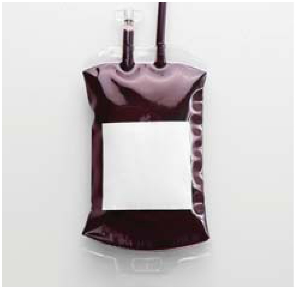
Image: Fresh whole blood remains a mainstay of treatment for haemophilia in Nigeria, although the country’s blood transfusion service is underdeveloped and donor screening is limited
© Shutterstock
Haemophilia A is an X-linked recessive disorder characterised by deficiency of FVIII and recurrent bleeding episodes, which may occur spontaneously (in severe haemophilia: FVIII <1%) or be provoked by trauma (in non-severe haemophilia: FVIII>1%) [1]. Haemarthrosis and arthropathy are common clinical features due to the role of synovial cells and chondrocytes as producers of the tissue factor pathway inhibitor [2,3]. However, no tissue is exempted from haemophilic bleeding diathesis [4]. Moreover, locally endemic parasitic diseases such as urinary schistosomiasis and intestinal helminthiasis are important causes of haematuria, haematochezia and iron deficiency among haemophiliacs living in tropical countries such as Nigeria [5, 6, 7].
Lack of FVIII concentrates is the single most important cause of high infant and childhood mortality among haemophiliacs in low-resource tropical countries [8]. Consequently, multiple transfusions with fresh whole blood (FWB) and blood products such as fresh frozen plasma (FFP) and cryoprecipitate (CP) are the main therapeutic options in Nigeria [9]. However, transfusion safety is poor because transmissible infections are prevalent, donor blood supply is scarce and screening facilities are inadequate [10,11]. Recent studies indicate that up to 15-20% of apparently healthy donors in Nigeria were infected with one or more transfusion transmissible viruses, including hepatitis B virus (HBV), hepatitis C virus (HCV) and human immune deficiency virus (HIV) [12,13]. The prevalence rates of individual transfusion transmissible viral infections (TTVIs) among blood donors in Nigeria are up to 4.111.1% for HBV, 1.8-3.6% for HCV, and 1.4-5.2% for HIV-1, while HIV-2 is relatively rare [12, 13, 14].
Image: For many people with haemophilia in Nigeria, first-line treatment is accessed through sub-tertiary hospitals with limited resources. Tertiary hospitals are generally located in cities, expensive and less accessible to patients and their carers
© Shutterstock

Donor screening is rudimentary in Nigeria, as the national blood transfusion service is grossly underdeveloped and responsibility for blood procurement, storage and safety largely rests on individual hospital blood banks [15]. The only viruses routinely tested for in blood banks are HIV, HBV and HCV, and the practice and quality of screening techniques have remained largely unchanged since the introduction of rapid immunoassay tests. Nigerian tertiary hospital blood banks have only a basic capacity to screen donors for TTVIs, and use rapid qualitative immunochromatographic kits in the majority of cases and, to lesser extent, standard incubation enzyme-linked immunosorbent assay (ELISA) techniques [15,16]. Immunoblot assays (for confirmation of reactive immunoassays) and window period tests (for confirmation of non-reactive assays) are not routinely available [15,16]. Nigerian sub-tertiary hospitals often lack adequate laboratory facilities and/or skilled manpower to conduct even basic immunoassays and serological tests to screen donor blood for TTVIs [15].
Sub-tertiary hospitals are more widely distributed (in both rural and urban areas) and cheaper than tertiary hospitals, and more accessible to haemophiliacs and their parents. Consequently, sub-tertiary hospitals are usually the first line of haemophilia care; the only therapeutic option for haemophiliacs (and indeed any bleeding patients), however, is the transfusion of FWB, which is often inadequately screened for TTVIs. As a therapeutic source of FVIII, FWB is only marginally effective: it has very high volume-to-efficacy ratio and may thus necessitate multiple transfusions (hence, multiple donor exposures) to achieve effective haemostasis. The FWB transfusion therapy offered to haemophiliacs in sub-tertiary hospitals may therefore be both microbiologically unsafe and therapeutically inefficient.
Haemophiliacs who survive eventually pass through the chain of medical referrals from sub-tertiary to tertiary hospitals. The tertiary hospitals are almost exclusively located in cities, and are thus expensive and not easily accessible to patients and their parents/ guardians. Although tertiary hospitals have the capacity to produce more haemostatically effective blood products, such as FFP and CP, FWB remains the most frequently transfused blood product in managing haemophilia for two reasons. First, production of FWB is technically simple and requires neither product separation nor sub-zero deep-freezing storage facilities as opposed to production of FFP and CP, which require greater technical skills and facilities for cold centrifugation, product separation and sub-zero storage [9]. Second, many haemophiliacs in Nigeria also suffer from chronic anaemia as a result of the combined effects of poverty, malnutrition, frequent haemophilic bleeding, and comorbid endemic tropical parasitic infestations that increase blood loss and cause iron deficiency [5, 6, 7]. Hence, the ease of production of FWB and its dual advantage as a product that can simultaneously stop active bleeding (due to its FVIII component) and treat anaemia (due to its red cell component) makes FWB the most frequently transfused product in the management of haemophilia in Nigeria [9].
The tertiary hospitals derive their blood products from donors who have screened negative (by basic immunoassay and serology, but not routinely tested for window period) for TTVIs. Therefore, in comparison with sub-tertiary hospitals, tertiary hospital blood products are safer – but only relatively so, as the absence of window period testing implies a significant residual risk of TTVIs [17]. By the time haemophiliacs access tertiary hospitals, they are likely to have had multiple transfusions with inadequately screened FWB administered at local sub-tertiary hospitals.
Against this background, we predicted that the prevalence of TTVIs would be high among haemophiliacs in Nigeria. To the best of our knowledge, the prevalence and pattern of TTVIs in haemophiliacs have not been previously studied and reported in Nigerian haemophiliacs. To investigate our prediction, we conducted a retrospective analysis of the prevalence and pattern of TTVIs (including HIV, HBV and HCV infections) among newly referred paediatric haemophiliacs as seen at the time of initial clinical evaluations in five tertiary hospitals in northern Nigeria. The clinical implications of our findings were discussed within the context of haemophilia.
Materials and methods
Clinical setting and study description
As in other developing countries, there is currently no organised system for the early detection, diagnosis, reporting, documentation or treatment of haemophilia in Nigeria [18]. Haemophilic bleeding diathesis is therefore often first reported to local sub-tertiary hospitals, where multiple transfusions with FWB is the only therapeutic modality for haemophilia. As treatment outcome becomes less satisfactory, patients are eventually referred to tertiary hospitals, where more effective therapeutic blood products such as FFP and CP may be available.
In view of the high infection risk associated with previous transfusions at sub-tertiary hospitals, haemophiliacs who arrived at our tertiary hospitals were regularly screened for TTVIs (including HIV, HBV and HCV) after confirmation of their diagnosis with FVIII assays. This is a retrospective cohort study of such data accrued from haemophilia patients who were screened for HIV, HBV and HCV infections as seen at different time intervals in five northern Nigerian tertiary hospitals: the University of Maiduguri Teaching Hospital, Maiduguri, North East Nigeria (1997-2007); State Specialist Hospital, Maiduguri, North East Nigeria (1997-2007); Federal Medical Centre, Birnin Kudu, North West Nigeria (2004-2008); Murtala Muhammad Specialist Hospital, Kano, North West Nigeria (2008-2011); and Aminu Kano Teaching Hospital, Kano, North West Nigeria (2008-2012). This study was conducted with the approval of local institutional ethics committees.
Selection of patients
Haemophilia patients who had received one or more transfusions with FWB at sub-tertiary hospitals were included in this study. Patients without a history of transfusion were excluded.
Haemophilia diagnosis and screening for transfusion transmissible infections
The patients studied were confirmed cases of haemophilia A, diagnosed on the basis of characteristic clinical profiles with low FVIII levels as assayed by automated coagulometers or by the one-stage manual assay technique [19]. Patients were categorised as severe (FVIII level <1%), moderate (FVIII level 1-5%) or mild (FVIII level >5%) haemophiliacs [19]. Each patient’s serum or plasma was screened for the surface antigen of HBV (HBsAg) and antibodies to HIV-1, HIV-2 and HCV using a combination of commercial rapid qualitative immunochromatographic test kits and ELISA techniques, in accordance with manufacturers’ guidelines.
Retrospective appraisal of number of previous transfusions
The number of previous FWB transfusions as documented by the referring sub-tertiary hospitals and/or as corroborated/narrated by patients’ parents/ guardians at the time of clinical history taking were recorded and collated for each patient.
Statistical data analysis
Data accrued from the five tertiary hospitals were collated and analysed. Prevalence rates of infections were expressed as percentages. Comparisons of parameters (age, disease severity and number of previous transfusions per patient) between patients with and without TTVIs were performed using Student’s t-test for mean values and Fisher’s exact test for percentages, with p-values of less than 0.05 taken as significant. Statistical analysis was performed using SPSS software, version 15.0 (SPSS Inc., Chicago, IL, USA).
Results
A total of 99 male haemophiliacs, aged 3.5–11.5 years, referred from sub-tertiary hospitals were seen during the period under review in the five tertiary hospitals. Two patients were excluded because of a lack of positive history of blood transfusion at the referring hospitals.
Of the 97 patients with positive history of blood transfusions, 24 (24.7%) had one or more TTVIs. The pattern and frequencies of TTVIs are shown in Table 1: HBV infection in 10 (41.7%) patients, HIV-1 infection in five (20.8%), HCV infection in four (16.7%), HBV and HIV co-infection in three (12.5%), and HBV and HCV co-infection in two (8.3%). HIV-2 infection was not found in any patient and no patients had triple co-infection with HIV, HBV and HCV.
Table 1
Pattern and frequencies of infections among 24 paediatric haemophiliacs with transfusion transmissible viral infections (TTVIs)
| TTVI | NUMBER OF PATIENTS INFECTED (%) |
|---|---|
| HBV | 10 (41.7) |
| HIV-1 | 5 (20.8) |
| HCV | 4 (16.7) |
| HBV and HIV-1 | 3 (12.5) |
| HBV and HCV | 2 (8.3) |
| All infections | 24 (100) |
The mean age, pattern of disease severity and number of transfusions per patient among haemophiliacs with and without TTVIs are shown in Table 2. In comparison with haemophiliacs without TTVIs, haemophiliacs with TTVIs had a significantly lower mean age (4.9 vs. 7.8; p=0.007), higher proportion of severe disease (62.5% vs. 26%; p=0.009), and higher mean number of FWB transfusions per patient (27.5 vs. 15.3; p=0.006).
Table 2
Age, disease severity and number of previous transfusions among 97 haemophiliacs with and without transfusion transmissible viral infections (TTVIs)
Discussion
Similar to other developing countries, the exact prevalence and incidence of haemophilia in Nigeria are currently unknown due to under-diagnosis, under-documentation and under-reporting of cases [18], coupled with high and early childhood mortality resulting from poor management [8]. As a result, this study is based on a relatively low sample size, despite a fairly extended period of review and the use of a multicentre approach. Nonetheless, Nigeria has the largest population in Africa and presumably carries the heaviest burden of persons living with haemophilia in Africa.
There is currently no organised system for the surveillance, early detection, diagnosis and treatment of haemophilia or other bleeding disorders in Nigeria. Consequently, haemophilic bleeding diathesis is often first reported to the cheaper and more accessible local sub-tertiary hospitals, which do not have the capacity to diagnose bleeding disorders and often lack adequate facilities and skills for basic immunoassays and serological screening of blood donors for TTVIs [15]. Sub-tertiary hospitals often empirically, but quite logically, treat any bleeding patient (including haemophiliacs) with multiple transfusions of FWB as the only therapeutic option at their disposal. It is only when the treatment outcome becomes unsatisfactory and the patient’s parents/guardians are financially capable that haemophiliacs are eventually referred to tertiary hospitals, where accurate diagnosis can be made and more effective therapeutic interventions such as FFP or CP may be offered.
The results of this study reveal that almost one quarter (24.7%) of haemophilia patients involved were infected with one or more TTVIs by the time they attended a tertiary hospital, after receiving multiple FWB transfusions at local sub-tertiary hospitals. The pattern and frequency of TTVIs among the infected haemophiliacs in this study reveals the predominance of HBV, followed by HIV and HCV, with relatively few cases of dual co-infections due to HBV and HIV or HCV. This pattern is consistent with the prevalence of individual TTVIs among blood donors in Nigeria [12,13]. It is also consistent with the pattern of endemicity of TTVIs in the general Nigerian population, wherein previous studies have reported the prevalence of TTVIs to be as high as 14.5% for HBV [20], 5.8%for HCV [21], and 3.4% for HIV [22].
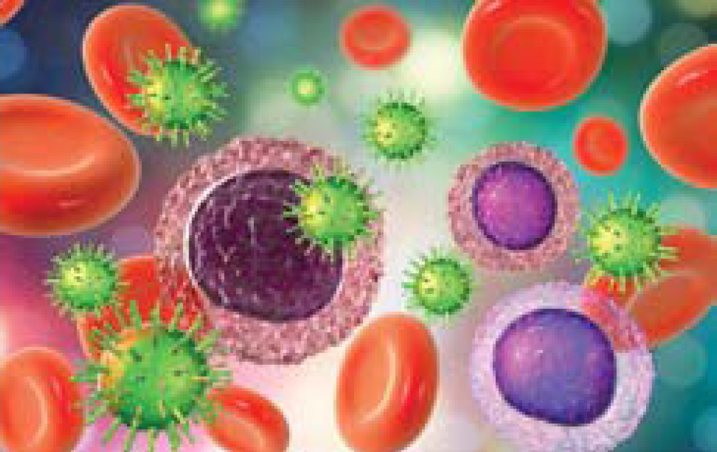
Image: A significant number of paediatric haemophiliacs in Nigeria are already infected with one or more transfusion transmissible viral infections by the time they attend a tertiary hospital, after receiving multiple blood transfusions at sub-tertiary hospitals
© Shutterstock
The high prevalence of TTVIs in the subjects of this study (24.7%) confirms our prediction that recurrent exposure to inadequately screened donor blood at sub-tertiary hospitals would lead to high prevalence of TTVIs in haemophiliacs. This is further supported by data showing that, in comparison with haemophiliacs without TTVIs, haemophiliacs with TTVIs had a higher prevalence of severe disease (implying a higher bleeding rate), with a commensurate higher mean number of transfusions per patient (implying greater exposure to infected donor blood). Moreover, in comparison with haemophiliacs without TTVIs, haemophiliacs with TTVIs were younger because they had higher prevalence of severe disease, an important predictor of early clinical presentation resulting in early referral to tertiary hospitals [4]. Hence, the findings of this study suggest that the risk of TTVIs in haemophilia is correlated with severe disease and the number of previous multiple blood transfusions received by the patients.
There is no doubt that a shift of clinical care from sub-tertiary to tertiary hospitals (with relatively better donor screening capabilities) would considerably reduce the risk of TTVIs for haemophiliacs in Nigeria. Nonetheless, in a tropical setting such as Nigeria, a significant residual risk of TTVIs would persist even in the tertiary hospitals, for several reasons. First, the routine immunoassay techniques for screening blood donors for HBsAg in Nigeria are basic rapid immunochromatographic tests [15]. These may not detect low levels of HBsAg in donors with occult HBV infection – unfortunately, nucleic acid tests for detecting occult HBV infections [23] are not routinely available in Nigeria. Second, the routine serological techniques for screening blood donors for HIV and HCV antibodies in Nigeria also employ rapid immunochromatographic kits [15]. These are relatively cheap but less accurate than standard incubation ELISA [24,25], which are used infrequently in Nigerian blood banks as they are expensive [25]. Third, more advanced procedures for the detection of window period infections ,such as p24 antigen for HIV [16], viral core antigen for HCV [26] and nucleic acid tests for both HIV and HCV [26,27], are not routinely available in Nigeria. Finally, there is a complete absence of viral inactivation technology for the elimination of the residual risks of TTVIs in blood and blood products in Nigeria [28].
For these reasons, we infer that the prevalence of TTVIs among haemophiliacs in Nigeria will continue to rise longitudinally (beyond the initial 24.7% found in this study) in direct proportion to the degree of multiple transfusions and cumulative donor blood exposure at any given time in the life of haemophilia patients. The strength of our inference is based on the findings of similar studies from other developing countries. The prevalence of TTVIs among multi-transfused haemophiliacs is reported to be as high as 17.3-47.5% in some Middle Eastern and North African countries [29,30], while a staggering prevalence of more than 50% was reported in Pakistan in the Asian subregion [31]. It can therefore be surmised that, apart from lack of FVIII concentrates, TTVIs are probably the greatest threat to healthy living and long-term survival among haemophiliacs living in poor and underdeveloped countries.
The acquisition of TTVIs has highly adverse clinical implications within the context of a pre-existing bleeding disorder such as haemophilia. The hepatitis viruses HBV and HCV often cause chronic liver disease (CLD) and hepatocellular cancer, causing significant damage to hepatocytes [32], the main producers of all coagulation factors with the exception of FVIII and von Willebrand factor (vWF) [33]. CLD would essentially ‘transform’ haemophilia from a ‘single-factor’ deficiency disorder to a ‘multiple-factor’ deficiency disorder. HIV is also inimical to haemophilia and, in addition to causing AIDS with recurrent opportunistic infections, can cause thrombocytopenia, jeopardising primary haemostasis function [34]. Hence, the haemostatic defects acquired due to viral CLD or HIV infection can aggravate the pre-existing haemophilic bleeding tendency, increase bleeding rates and worsen the prognosis of infected haemophiliacs. TTVIs are particularly undesirable in haemophiliacs in countries such as Nigeria, as they would invariably worsen the prognosis of an already under-treated inherited bleeding disorder.
The risk of acquiring TTVIs among haemophiliacs in Nigeria, and indeed other developing countries, can only be significantly reduced by upgrading the national transfusion service and blood safety protocols. This must include efficient donor screening procedures, effective viral inactivation techniques, and the production or regular provision of recombinant blood products, including FVIII concentrates, to a level whereby they are as obtainable as in (developed) countries where TTVIs among haemophiliacs have become history [35]. Attaining this haemophilia healthcare ‘utopia’ requires a great deal of political will and national economic development, which for tropical Africa is likely only to be achieved in the distant future.
In the meantime, healthcare personnel working in low-resource tropical areas with little or no access to FVIII concentrates, can minimise multiple blood transfusions and reduce the risk of TTVIs in the management of haemophiliacs in two ways. First, healthcare workers in tropical regions should incorporate regular screening and treatment for common haemorrhagic parasitic diseases and iron supplementation into the standard of care for haemophiliacs. This strategy will reduce gastrointestinal and urinary blood loss, prevent iron deficiency and decrease the incidence of anaemia, thereby cutting down the need for multiple transfusion and diminishing the risk of acquiring TTVIs among haemophiliacs. This recommendation has been made in previous studies based on tropical clinical experience [5,6]. Second, the lack of adequate use of pharmacological agents such as desmopressin and antifibrinolytic agents (epsilon amino caproic acid and tranexamic acid) was identified in a previous study as a major limitation in the management of haemophilia in tropical developing countries [8]. Desmopressin raises vWF and FVIII levels, while antifibrinolytic agents inhibit fibrin clot degradation; all have been shown to be useful in controlling haemophilic bleeding, especially in patients with mild and moderate haemophilia [36,37]. Tranexamic acid in particular has been widely and successfully used alone or in combination with other anti-haemophilic agents in the prevention and management of bleeding due to accidental or surgical trauma in haemophilia patients [37]. The only exception to the use of anti-fibrinolytic agents is bleeding in the urinary tract, because of the possible risk of fibrin clot obstructive nephropathy [37]. We therefore strongly recommend that haemophilia healthcare givers in tropical regions should intensify the use of these pharmacological agents (since they are cheaper and more widely available than FVIII concentrates) as a way of minimising multiple blood transfusions and reducing the risk of TTVIs.
Conclusion and recommendations
This study has limitations which mean that the conclusions we have been able to draw are necessarily broad. The retrieval and acquisition of retrospective data is particularly difficult in the Nigerian setting due to poor record-keeping and documentation. Our data set is relatively small and we recognise that there may be a clustering or grouping of prevalence data within the individual sites involved in the study and/or over different time periods. We were unable to stratify our findings on a site-by-site basis, however, as the total number of eligible patients in the study is quite small due to the low survival rate of haemophiliacs in Nigeria. A larger cohort may also have detected triple infection with HIV, HBV and HCV.
We can, however, conclude that the prevalence of TTVIs among paediatric haemophiliacs in Nigeria is high. The risk is correlated with disease severity and the number of previous blood transfusions, within the context of a lack of access to FVIII concentrates, an underdeveloped transfusion service, inadequate donor screening and poor transfusion safety.
Our intention is to offer solutions to the problem of TTVIs in the Nigerian haemophiliacs by recommending ways in which the need for transfusion can be minimised. There is a need for the Nigerian government to upgrade the national blood transfusion service and set up standard haemophilia care centres with an adequate supply of FVIII concentrates for regular prophylaxis and treatment. The practice of offering regular screening and treatment for common haemorrhagic parasitic diseases, iron supplementation, and adopting the use of pharmacological agents in the standard of care for haemophilia can help to reduce the need for quite so many multiple blood transfusions, and therefore help to minimise exposure to infected donor blood. Together, these recommendations can start to address the issues caused by the prevalence of TTVIs in haemophiliacs and other transfusion-dependent patient groups in Nigeria – and, indeed, other underdeveloped countries.